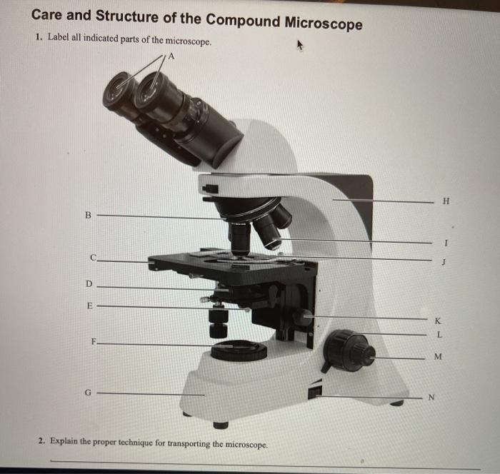
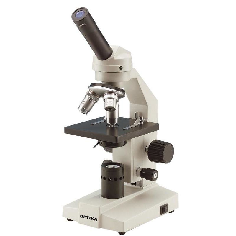
38 label the indicated parts of the microscope
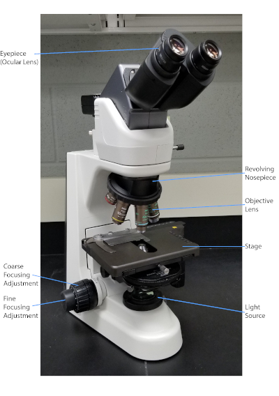
07-894-5866 | 1 ml, 25/Pkg | TruCan™ DAPPi+L4 - Patterson Vet 03.10.2013 · Volume: 1 ml Package Quantity: 25/Pkg Presentation: Modified Live Virus in a Powder Cake with Killed Virus and Inactivated Bacterin as a Sterile Diluent Doses Per Unit: 25 Container Type: Vial Delivery Type: Injection Reconstitution Required: Y Biological Type: Modified Live Diseases Indicated For: Canine Distemper, Infectious Canine Hepatitis, Canine … Solved Care and Structure of the Compound Microscope 1. - Chegg Expert Answer 100% (1 rating) Answer 1) A. Ocular lens. B. Rotating nose piece. C. Stage. D. Condensor. E. Iris diaphragm. F. Light source. G. Base. H. Arm. I. Objective lens. J. Mechanical stage. K. Condensor knob. L. Coarse adjustment. M. Fine adjustment. N. Light control. Micr … View the full answer
› articles › s41586/022/04977-7Formation of moiré interlayer excitons in space and time | Nature Aug 17, 2022 · c, The heterostructure can be identified in the real-space mode of the microscope. The WSe 2 /MoS 2 and WSe 2 regions of interest are indicated by red and orange circles, respectively (10-µm ...

Label the indicated parts of the microscope
Tri-Heart® Plus Chewable Tablets - Patterson Vet Overview (Ivermectin, Pyrantel) Indicated to prevent canine heartworm disease by eliminating the tissue stage of heartworm larvae (drofilaria immitis) for a month after infection and for the treatment and control of ascarids (toxocara canis, toxascaris leonina) and hookworms (ancylostoma caninum, uncinaria stenocephala, ancylostoma braziliense). The Microscope - City University of New York Care and Structure of the Compound Microscope I. label alt indicated parts of the microscope oos¿piece 2. the proper techntque for transporting the microscope one hand arm . 33 . Review Sheet 3 34 3. Each of the following statements IS either true or false If true, write T on the answer blank If false, correct the statement by Label the microscope — Science Learning Hub Use this interactive to identify and label the main parts of a microscope. Drag and drop the text labels onto the microscope diagram. eye piece lens diaphragm or iris coarse focus adjustment stage base fine focus adjustment light source high-power objective Download Exercise Tweet
Label the indicated parts of the microscope. EXERCISE 3 Parts of the microscope Flashcards | Quizlet vertical bar used to grasp and carry the microscope base lower portion of the microscope coarse adjustment knob large knob on the lower arm used to achieve initial focus condenser lens below the stage that produces a cone of light which is focused on the object; a knob raises and lowers the condenser to adjust the brightness of the field auctions.dreweatts.com › past-auctions › drewea1A FINE GEORGE II MAHOGANY CASED CUFF PATTERN MONOCULAR MICROSCOPE A FINE GEORGE II MAHOGANY CASED CUFF PATTERN MONOCULAR MICROSCOPEJOHN CUFF, LONDON, MID 18th CENTURYThe body tube with stepped moulded shuttered eyepiece over ogee waist and objective tube incorporating marks for six positions on an exponential scale numbered 1 to 6, supported via a tapered collar set in a ring attached to a vertical slide moving against the fixed limb upright marked with six ... Solved Lab 1- The Microscope-Lab Report 1. Using the - Chegg Science Biology Biology questions and answers Lab 1- The Microscope-Lab Report 1. Using the picture, label the indicated parts of the microscope with the terms listed in the blue box. Each term is used only once. (Note: 1, 4, and 5 are intentionally not included.) 2. 3. 6. 7. 8. 9. 10. 11. 12. 13. 14. a. Objectives b. Mechanical stage c. Arm d. Microscope World | Shop Microscopes For Every Application Labeling the Parts of the Microscope This activity has been designed for use in homes and schools. Each microscope layout (both blank and the version with answers) are available as PDF downloads. You can view a more in-depth review of each part of the microscope here. Download the Label the Parts of the Microscope PDF printable version here.
Answered: SOMATIC CELL DIVIS SPECIES, EA CH WITH… | bartleby Images The images on the left represent events of a mitotic division in a haploid cell in which N=2. In the other boxes in each row, draw the same events for cells with the indicated chromosome number. SPECIES A Haploid, N = 2 SPECIES B Haploid, N = 3 SPECIES C Diploid, N = 2 SPECIES D Diploid, N = 3 NAME THE PHASE LABEL: cytoplasm núclear ... Label parts on microscope Flashcards | Quizlet Body Tube on microscope High power objective Low power objective Stage Opening Diaphragm Lever Light on microscope Coarse adjustment Fine adjustment Stage Clip Stage on microscope Base Sets found in the same folder Cell Organelles Biology Honors- Characteristics of Life… WHMIS Symbols 9 terms jcacorn A FINE GEORGE II MAHOGANY CASED CUFF PATTERN MONOCULAR MICROSCOPE It is entirely the responsibility of the buyer to acquaint himself with the precise EURO to UK Sterling exchange rate on the day of the sale in this regard, and the auctioneer accepts no responsibility whatsoever if the qualifying rate is different to the rate indicated. All items in this catalogue that are marked with λ are potentially qualifying items, and the royalty charge will be … Iodine Test: Definition, Principle, Results I ResearchTweet 21.04.2022 · Label two test tubes with the words “test sample” and “control sample.” 2. In a clean and dried test tube designated as a test sample, take a small sample (solid sample: 500 mg-1000 mg; liquid sample: 1 ml). 3. In the clean and dried test tube designated as the control sample, pour 1 mL of purified water. 4. To both test tubes, add 2-3 drops of Lugol’s iodine solution and …
Using the Microscope: Basic Tutorial: Part 2: Components. - Micrographia The Eye. The cornea and the eye lens are the final optical components in the image-forming path to the retina. In a person with normal vision, the eyelens will be relaxed as though the eye is forming an image of a very distant object, and the focusing controls on the microscope used to achieve image sharpness. SOLVED:Use the information below to label the parts of the microscope ... Let's get started. A So, for a we have the outside of the eye of a white region. This is Venus Clara. Well, B next, we have the the back of the eye for region that detects light. This is for retina. See, this is pointing to the vessels and coming into the eye, but provides blood. So is so is D. And if you look very closely see is pointing for ... Label The Microscope Parts! Diagram | Quizlet Label The Microscope Parts! + − Flashcards Learn Test Match Created by awesome3wash Terms in this set (16) Body Tube Connects the eyepiece to the objective lenses Revolving Nose Piece holds the objectives and can be easily rotated to change viewing power. Scanning Objective City University of New York The Microscope EXERCSE Mar; qldb Lab Time/Date Name Care and Structure of the Compound Microscope I. Label all indicated parts of the microscope. esQi€ce 2. Explain the proper technique for transporting the microscope. 0b qs Ont en arm loose. 33 . 34 Review Sheet 3 3. Each of the following statements is either true or false.
This is a common compound microscope. Label its parts from A ...
pypi.org › project › connected-components-3dconnected-components-3d · PyPI Aug 18, 2022 · cc3d: Connected Components on Multilabel 3D Images. Fig. 1. Binary and Multilabel Connected Components Labeling (CCL) 2D images are shown for simplicity. (a) A binary image (foreground white, background black) (b) 4-connected CCL of binary image (c) 8-connected CCL of binary image (d) A multilabel image (e) 4-connected CCL of multilabel image (f) 8-connected CCL of multilabel image

0714nature test worksheet
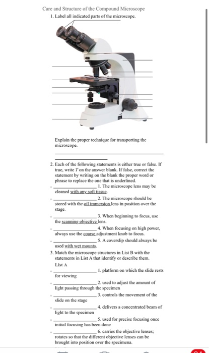
Activity 3.1.docx - Denise Learned Bio201 The Microscope... Denise Learned Bio201 The Microscope Care and Structure of the Compound Microscope 1. Label all indicated parts of the microscope. 2. Explain the proper technique for transporting the microscope. Answer: HOLD IT IN AN UPRIGHT POSITION WITH ONE HAND ON ITS ARM AND THE OTHER SUPPORTING ITS BASE

Compound Microscope Parts – Labeled Diagram and their ...
PDF Parts of the compound microscope: Write the correct label for each part ... base and arm a. eyepiece, what you look in to see an image 14. coarse focus knob b. used to produce a more magnified image 15. stage clip c. used to bring the image into sharp focus 16. stage d. flat surface on which slide is placed 17. fine focus knob e. secures slide in place before viewing 18. high power objective …

Labeling the Parts of the Microscope | Microscope activity ...
microscope worksheet pp with answers - Worksheet: The... ____________________ _________________________________________________part function ocular eyepieces to look thru; magnify 10 times arm hold it while carrying 'scope coarse adjustment knob fine adjustment knobbase light switch substage light condenser iris diaphragm lever mechanical stage stage for coarse focusing for fine focusing 'scope sits on …
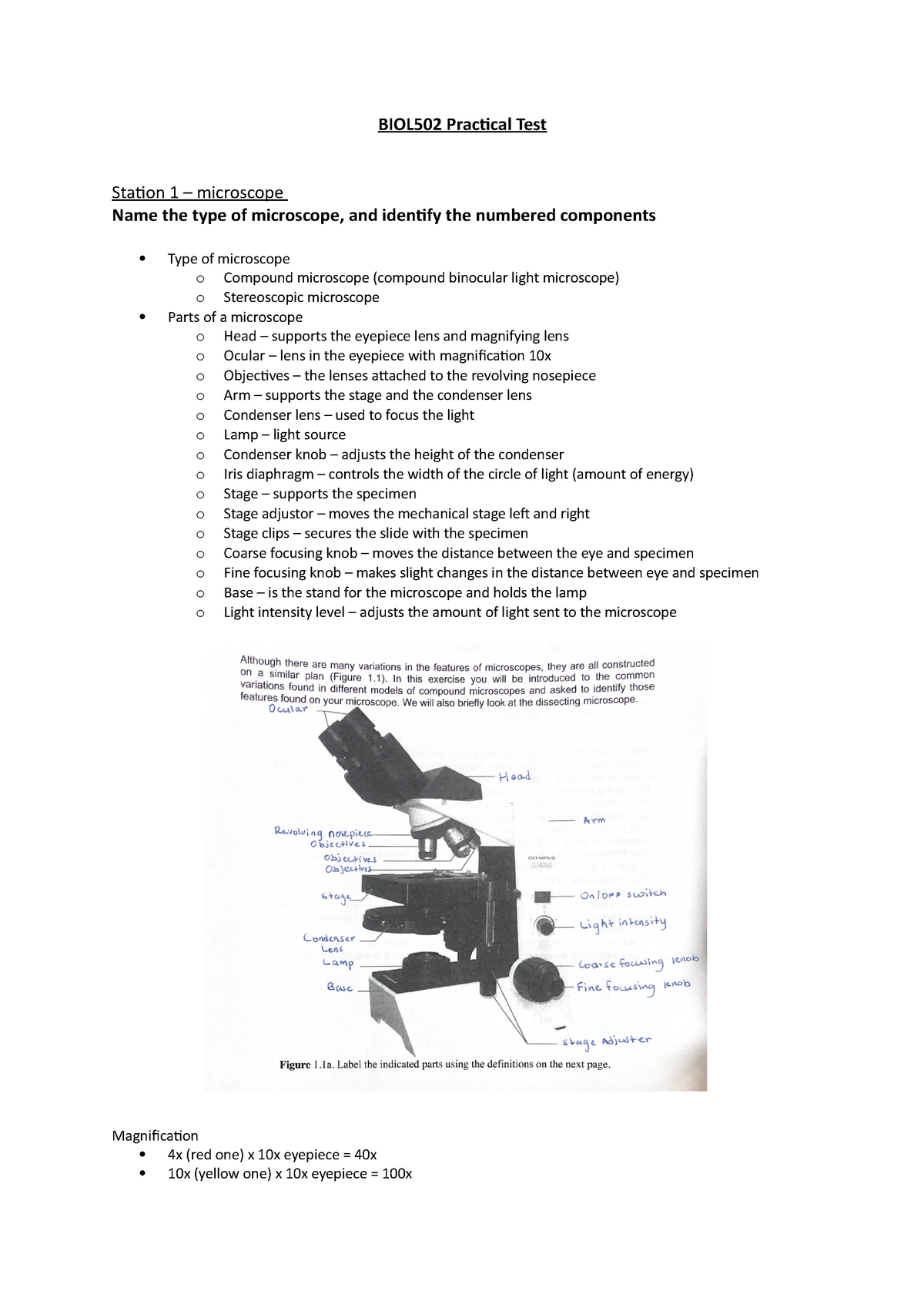
BIOL502 Practical Test - BIOL502 Practical Test Station 1 ...
microbenotes.com › parts-of-a-microscopeParts of a microscope with functions and labeled diagram Sep 17, 2022 · Figure: Diagram of parts of a microscope. There are three structural parts of the microscope i.e. head, base, and arm. Head – This is also known as the body. It carries the optical parts in the upper part of the microscope. Base – It acts as microscopes support. It also carries microscopic illuminators.

Lab Book - RS 3.pdf - NAME LAB 'I'IME'DATE The Microscope ...
Microscope Parts & Functions - AmScope Body: Often referred to as the head, the body is the upper part of a microscope including, eyepieces and objectives. Most modern microscopes are modular in the sense that the same body can be used with different bases and vice versa.
Solved Care and Structure of the Compound Microscope 1 ...
connected-components-3d · PyPI 18.08.2022 · This rendered these other packages too slow for my use case as it required masking each label and running the connected components algorithm once each time. For reference, there are often between hundreds to thousands of labels in a given volume. The benefit of this package is that it labels all connected components in one shot, improving performance by …
Labeling Microscope | Cell Structure Quiz - Quizizz
Virtual Microscope - NCBioNetwork.org Lesson Description BioNetwork’s Virtual Microscope is the first fully interactive 3D scope - it’s a great practice tool to prepare you for working in a science lab. Explore topics on usage, care, terminology and then interact with a fully functional, virtual microscope. When you are ready, challenge your knowledge in the testing section to see what you have learned.
Solved Care and Structure of the Compound Microscope 1 ...
www1.udel.edu › biology › ketchamUD Virtual Compound Microscope - University of Delaware ©University of Delaware. This work is licensed under a Creative Commons Attribution-NonCommercial-NoDerivs 2.5 License.Creative Commons Attribution-NonCommercial-NoDerivs 2
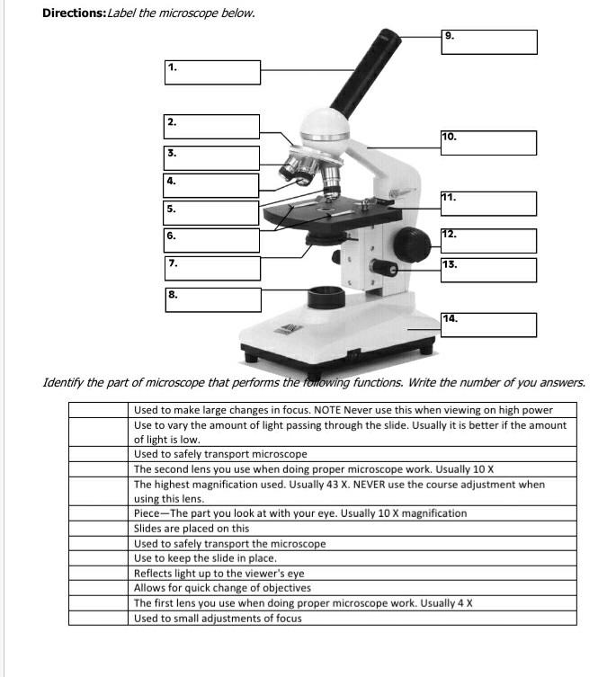
Solved Directions:Label the microscope below. 9. 1. 111 2 ...
LABEL ALL INDICATED PARTS OF THE MICROSCOPE.docx - Course Hero 4 LABEL ALL INDICATED PARTS OF THE MICROSCOPE. 5 LABEL ALL INDICATED PARTS OF THE MICROSCOPE. 6 EXPLAIN THE PROPER TECHNIQUE FOR TRANSPORTING THE MICROSCOPE. WHEN TRANSPORTING THE MICROSCOPE, HOLD IT IN AN UPRIGHT POSITION WITH ONE HAND ON ITS ARM AND THE OTHER SUPPORTING ITS BASE.
Microscope Quiz
Solved Care and Structure of the Compound Microscope 1. - Chegg Anatomy and Physiology. Anatomy and Physiology questions and answers. Care and Structure of the Compound Microscope 1. Label all indicated parts of the microscope. 2. Explain the proper technique for transporting the microscope. Question: Care and Structure of the Compound Microscope 1.
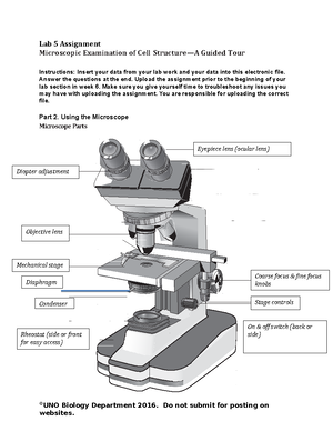
Lab 5 Assignment - Lab 5 Assignment Microscopic Examination ...
compound microscope parts (labeling) Flashcards | Quizlet what is 1? nosepiece (turret) - holds and spins the objective lenses what is 2? 4x objective lens - the 'scanning" objective lens with the lowest magnification what is 3? 10x objective lens - the "low" power objective lens what is 4? 40x objective lens - the "high" power objective lens with the most magnification what is 5?

Parts of a microscope with functions and labeled diagram
Parts of a microscope with functions and labeled diagram 19.04.2022 · Figure: Diagram of parts of a microscope. There are three structural parts of the microscope i.e. head, base, and arm. Head – This is also known as the body. It carries the optical parts in the upper part of the microscope. Base – It acts as microscopes support. It also carries microscopic illuminators.
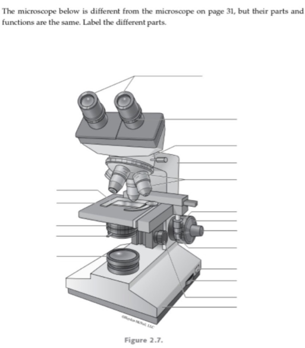
Solved The microscope below is different from the microscope ...
UD Virtual Compound Microscope - University of Delaware ©University of Delaware. This work is licensed under a Creative Commons Attribution-NonCommercial-NoDerivs 2.5 License.Creative Commons Attribution-NonCommercial-NoDerivs 2.5 License.
Microscopy and Staining Techniques in Bacteria | Microbiology ...
› 220824 › p12Techmeme: The Ethereum Foundation says it will begin the ... Aug 24, 2022 · The Ethereum Foundation says it will begin the Merge on September 6, split into two parts, the second running between September 10-20 — - The Bellatrix upgrade for The Merge is set for September 6, and Paris will follow several days later. — Node operators must download clients updates before the Bellatrix upgrade is activated.

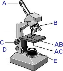
Label the parts of the microscope indicated by each letter ...
Microscope Parts and Functions Body tube (Head): The body tube connects the eyepiece to the objective lenses. Arm: The arm connects the body tube to the base of the microscope. Coarse adjustment: Brings the specimen into general focus. Fine adjustment: Fine tunes the focus and increases the detail of the specimen. Nosepiece: A rotating turret that houses the objective lenses.

Lab Chapter #2 - The Microscope Diagram | Quizlet
PDF The Microscope - Holly H. Nash-Rule, PhD After the parts of the microscope have been identified, turn on the light and adjust the interpupillary distance so that ... Care and Structure of the Compound Microscope 1. Label all indicated parts of the microscope. Ocular lenses Rotating nosepiece Objective lenses Stage Mechanical stage Iris diaphragm lever Condenser Substage light Head Arm

Parts of a Microscope - SmartSchool Systems
Review Sheet: the microscope Flashcards | Quizlet which part of the microscope: delivers a concentrated beam of light to the specimen condenser which part of the microscope is used for precise focusing once initial focusing has been done fine adjustment knob

Bobcat 220 Excavator PDF Service Manual
(Solved) - Care and Structure of the Compound Microscope 1. Label all ... 1 Answer to Care and Structure ...

Label the microscope — Science Learning Hub
NBC Sports | Live Streams, Video, News, Schedules, Scores and more Watch the NFL's Sunday Night Football, NASCAR, the NHL, Premier League and much more. Live stream, watch highlights, get scores, see schedules, check standings and fantasy news on NBCSports.com
Solved Care and Structure of the Compound Microscope 1 ...
Label the parts of the microscope below in the - Course Hero Identify each of the specimens in the indicated spaces below. Specimen 1 -Red blood cells Specimen 2 - House spider eyes Specimen 3 - Sleletal Muscle Specimen 4 - Human Hair. Specimen 5 - Immune system cells Specimen 6 - Insect spiracle Specimen 7 - Daisy anther Specimen 8 - Mold Specimen 9 - Bone cell Specimen 10 - House fly mouth Discussion ...

Semi-automatic Manual Marking Machine YL-360 Sign Nameplate Coding Machine Equipment Parameter Label Printer YZ
Parts of the Microscope with Labeling (also Free Printouts) Let us take a look at the different parts of microscopes and their respective functions. 1. Eyepiece it is the topmost part of the microscope. Through the eyepiece, you can visualize the object being studied. Its magnification capacity ranges between 10 and 15 times. 2. Body tube/Head It is the structure that connects the eyepiece to the lenses.

PPT - Label the parts on your microscope picture. PowerPoint ...
Label The Parts Of A Microscope Worksheet Answers Label the parts of the microscope indicated and state the. Can be used for practice or as a quiz. We tried to locate some good of microscope parts and use worksheet answer key along with labeling the parts of. Handphone Tablet Desktop Original Size There are many key components to understand when utilizing a microscope. Get thousands of teacher ...

Ch.3 Westcott Microbiology Flashcards | Quizlet
› iet › microscopeVirtual Microscope - NCBioNetwork.org Lesson Description BioNetwork’s Virtual Microscope is the first fully interactive 3D scope - it’s a great practice tool to prepare you for working in a science lab. Explore topics on usage, care, terminology and then interact with a fully functional, virtual microscope.

Basic Geology 1. Crystallography. - ppt download
Compound Microscope: Parts of Compound Microscope - BYJUS The parts of the compound microscope can be categorized into: Mechanical parts; Optical parts (A) Mechanical Parts of a Compound Microscope. 1. Foot or base. It is a U-shaped structure and supports the entire weight of the compound microscope. 2. Pillar. It is a vertical projection. This stands by resting on the base and supports the stage. 3. Arm

Microscope Diagram | Quizlet
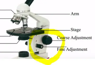
PDF Microscope Parts and Functions - WPMU DEV Storing The Microscope Using the microscope • Always observe using the LOWEST POWER objective first. • Focus using the COARSE ADJUSTMENT KNOB to bring the object into focus. Bring the object into sharp focus by using the fine adjustment knob. • Focus, and then move to a higher power objective, if needed.
Parts of the Microscope with Labeling (also Free Printouts ...
Label the microscope — Science Learning Hub Use this interactive to identify and label the main parts of a microscope. Drag and drop the text labels onto the microscope diagram. eye piece lens diaphragm or iris coarse focus adjustment stage base fine focus adjustment light source high-power objective Download Exercise Tweet

Parts of Microscope (Labeling) Diagram | Quizlet
The Microscope - City University of New York Care and Structure of the Compound Microscope I. label alt indicated parts of the microscope oos¿piece 2. the proper techntque for transporting the microscope one hand arm . 33 . Review Sheet 3 34 3. Each of the following statements IS either true or false If true, write T on the answer blank If false, correct the statement by
Solved Care and Structure of the Compound Microscope 1 ...
Tri-Heart® Plus Chewable Tablets - Patterson Vet Overview (Ivermectin, Pyrantel) Indicated to prevent canine heartworm disease by eliminating the tissue stage of heartworm larvae (drofilaria immitis) for a month after infection and for the treatment and control of ascarids (toxocara canis, toxascaris leonina) and hookworms (ancylostoma caninum, uncinaria stenocephala, ancylostoma braziliense).
Parts of a Microscope Quiz
Parts Of The Microscope Label Teaching Resources | TpT
Caterpillar cat d8 l track type tractor dozer bulldozer ...
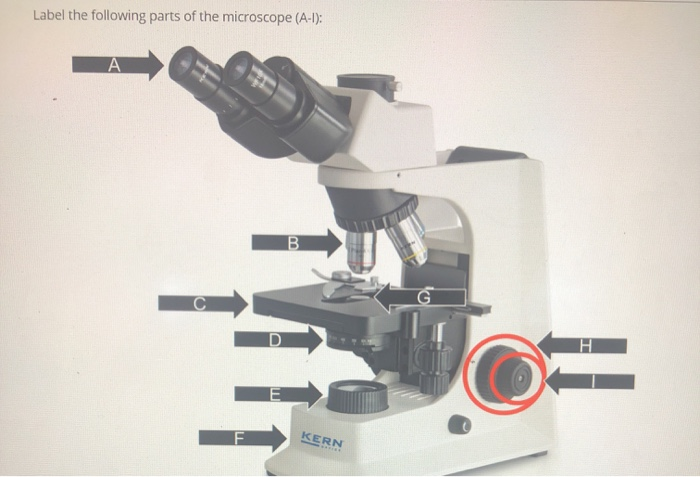
Solved Label the following parts of the microscope (A-1 ...

Label Microscope Diagram - EnchantedLearning.com
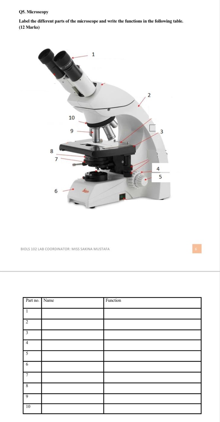
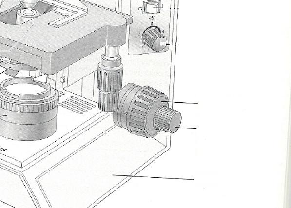
Solved Q5. Microscopy Label the different parts of the ...

The Microscope

Compound Microscope Parts – Labeled Diagram and their ...
Exercise 3: The Microscope Flashcards - Easy Notecards
Post a Comment for "38 label the indicated parts of the microscope"