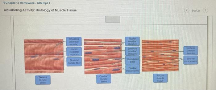
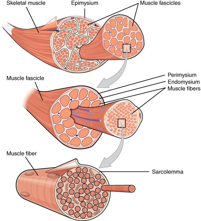
43 art-labeling activity: structure of muscle tissues
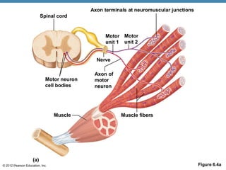
Chapter 3: Lab 3 Mastering Assignment Questions - Quizlet Cells join to form tissues, and the tissues then join to form the organs that work together in organ systems. ... When your biceps brachii (upper arm) muscle contracts, ultimately and most directly, what is producing the movement? 1. You move your arm. ... Art-labeling Activity: Figure 3.9. Drag the appropriate labels to their respective targets. BIOLOGY 2301 EXAM 1 - STUDY SET CHAPTER, 1, 2, 3, 4 Study with Quizlet and memorize flashcards containing terms like Identify the structure located within the mediastinum, The study of the liver is to gross anatomy as the study of a liver cell is to, Cardiovascular function is an example of and more. ... Components: skeletal muscles, cardiac muscle of the heart, and smooth muscles in the walls ...
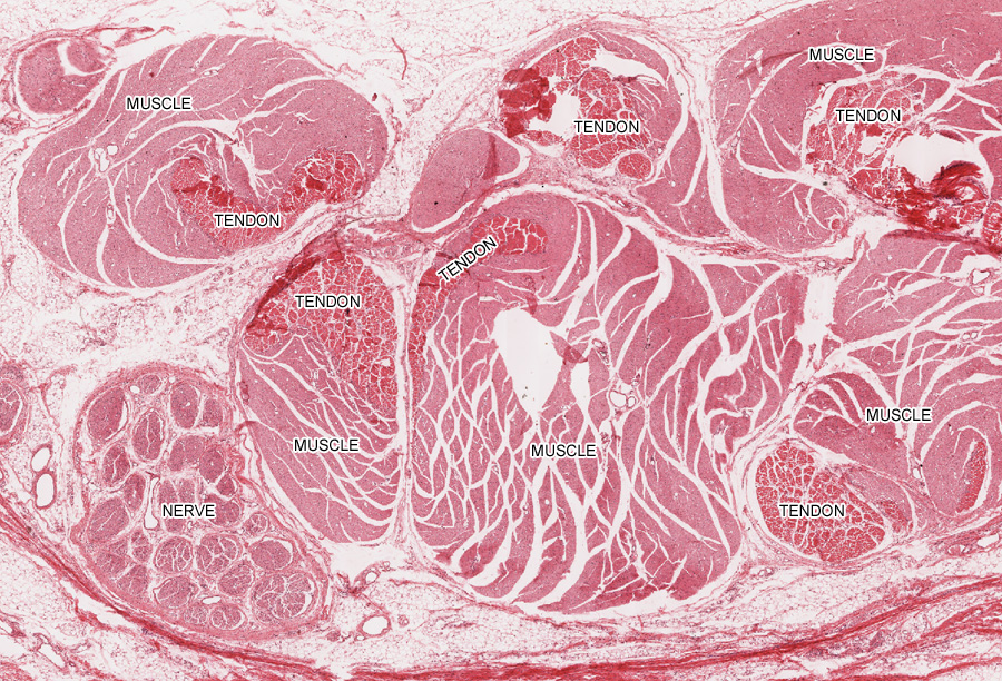
Solved Art-labeling Activity: Dense Connective Tissues 1547 | Chegg.com Transcribed image text: Art-labeling Activity: Dense Connective Tissues 1547 LOCATIONS mu andre w Dees . FUNCTIONS Tenson Denne Connective Fibro TIL II LOCATION மாயை | Dane Regular Connect Tour FUNCTIONS Colpen Geos ste ganun LOCATIONS Collagen கொயவரை பகாம்பாள் rude FUNCTIONS of LOCATIONS: Between skeletal musdes and skeleton (tendons and aponeuros ...
Art-labeling activity: structure of muscle tissues
[Solved] Art-Labeling Activity: | Course Hero All tutors are evaluated by Course Hero as an expert in their subject area. The image attached below is a sketch of the layers of the skin. There are 5 layers of the epidermis, which are categorized as follows (from superficial to deep): Stratum corneum: outer layer of the epidermis. Consists of dead (anuclear), keratin-filled cells. dokumen.pub › human-physiology-an-integratedHuman physiology : an integrated approach [Eighth edition ... Cell Junctions Hold Cells Together to Form Tissues Epithelia Provide Protection and Regulate Exchange Connective Tissues Provide Support and Barriers Muscle and Neural Tissues Are Excitable Tissue Remodeling Apoptosis Is a Tidy Form of Cell Death Stem Cells Can Create New Specialized Cells Emerging Concepts Induced Pluripotent Stems Cells Focus ... quizlet.com › 488100546 › chapter-3-lab-3-masteringChapter 3: Lab 3 Mastering Assignment Questions - Quizlet When your biceps brachii (upper arm) muscle contracts, ultimately and most directly, what is producing the movement? 1. You move your arm. 2. Proteins within the cells of the biceps brachii slide past each other lengthwise, shortening the muscle. 3. Your biceps brachii shortens. 4. Your brain tells your muscle to contract.
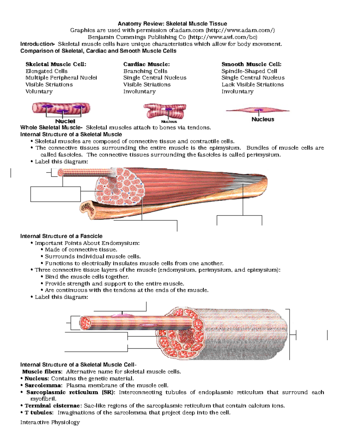
Art-labeling activity: structure of muscle tissues. art-labeling activity: sarcomere structure ... The structure of a skeletal muscle fiber Part A Drag the labels onto the diagram to identity structural features associated with a skeletal muscle fiber. Solved Lab 6 Muscular Tissue And System Art Labeling Chegg Com The sarcomere is the contraction unit in the skeletal muscles that are under the control of motor.. Sarcomeres are able to ... Tissues Lab p5-8.pdf - Reset Loose CT Hyaline cartilage... Correct The structure labeled D is a collagen fiber. Collagen fibers are thick, and appear pink due to staining. Art-Labeling Activity: Structure of muscle tissues Part A Drag the appropriate labels to their respective targets. Human physiology : an integrated approach [Eighth edition ... Muscle and Neural Tissues Are Excitable Tissue Remodeling ... Chemical Signals Influence Smooth Muscle Activity Cardiac Muscle CHAPTER SUMMARY REVIEW QUESTIONS CHAPTER 13: Integrative Physiology I: Control of Body Movement ... Selected figures from the text can be assigned as Art-Labeling Activities in Mastering AP. See p. 44 … Art-labeling Activity: The Structure of a Skeletal Muscle Fiber Start studying Art-labeling Activity: The Structure of a Skeletal Muscle Fiber. Learn vocabulary, terms, and more with flashcards, games, and other study tools.
Answer correct art based question chapter 4 question - Course Hero ANSWER: Correctmultinucleate cells branched cells intercalated discs situated between cells striations tendons and ligaments attached to bones heart ducts of certain glands dense irregular connective tissue smooth muscle tissue skeletal muscle tissue cardiac muscle tissue Campbell Biology, 12th Edition [12nbsped.] 9780135988046 35 Vascular Plant Structure, Growth, and Development Concept 35.1 Plants have a hierarchical organization consisting of organs, tissues, and cells Vascular Plant Organs: Roots, Stems, and Leaves Dermal, Vascular, and Ground Tissues Common Types of Plant Cells Concept 35.2 Different meristems generate new cells for primary and secondary growth chapter 9- Mastering A and P, Chapter 9-1 The muscular Tissue - Quizlet Art-labeling Activity: The structure of a skeletal muscle fiber PICTURE Chapter Test - Chapter 9 Question 3 Which thin-filament-associated structure is distinguished by its constituents of three globular subunits, one of which has a receptor that binds two calcium ions? a) G-actin b) nebulin c) tropomyosin d) troponin D ... Mastering AP Chapter 1 - The Human Body Flashcards | Quizlet It is the study of the structure of body parts and their relationships with one another. It is the study of all chemical reactions that occur within body cells. ... It is the study of tissues. and more. ... Art-labeling Activity: Figure 1.8. Art-labeling Activity: Figure 1.9. Moving from simpler to more complex, which level of organization is ...
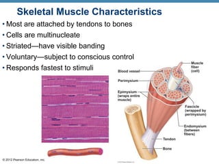
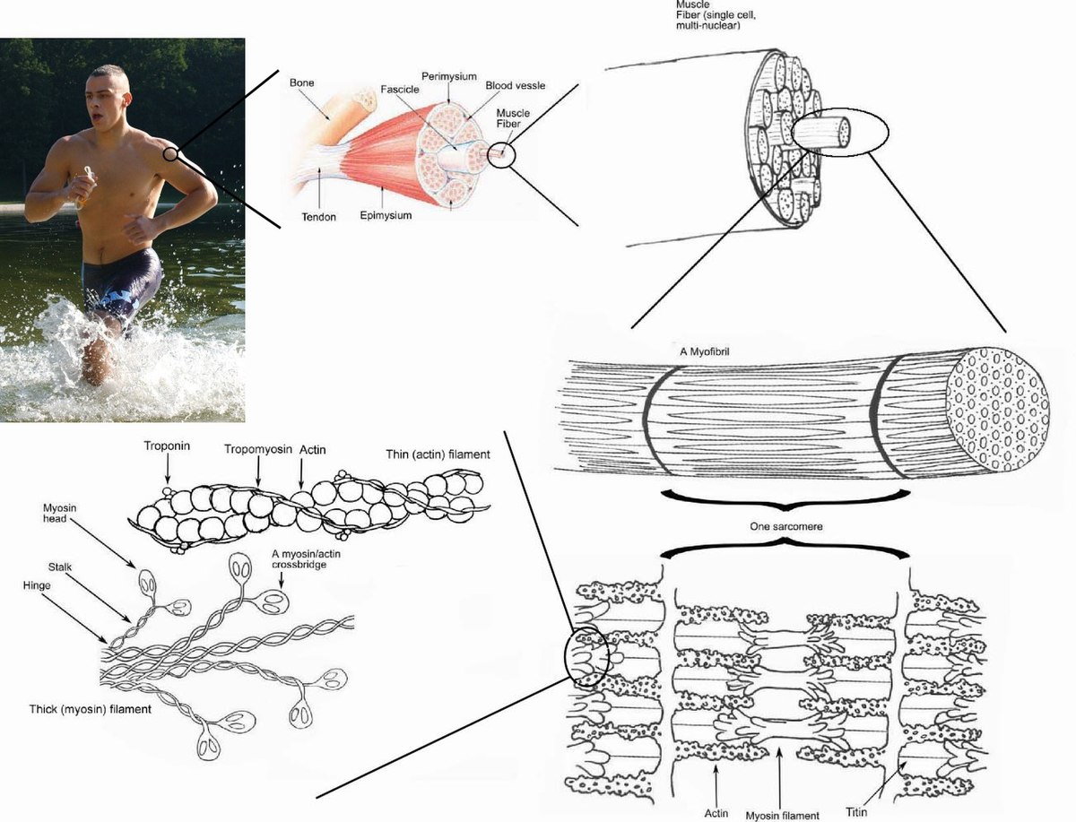
Art- labeling Activity Flashcards | Quizlet Study with Quizlet and memorize flashcards containing terms like , , and more. quizlet.com › 412756448 › mastering-ap-chapter-6Mastering AP Chapter 6 - Bones and Skeletal Tissues - Quizlet Study with Quizlet and memorize flashcards containing terms like Art-labeling Activity: Figure 6.2 Long bone Short bone Irregular bone Flat bone Sesamoid bone (short), Art-labeling Activity: Figure 6.4a Distal epiphysis Diaphysis Medullary cavity Compact bone Articular cartilage Proximal Epiphysis Spongy bone Epiphyseal line, Art-labeling Activity: Figure 6.4c Yellow bone marrow Nutrient ... art-labeling activity: sarcomere structure - 4-h-dairy-posters Each skeletal muscle has three layers of connective tissue that enclose it provide structure to the muscle and compartmentalize the muscle fibers within the muscle. Sarcomere Lower middle box Step-by-step explanation A band The thick myosin filaments are arranged across the myofibrils and the cell causing them to refract light and generate the ... [Solved] Art-Labeling Activity: | Course Hero Step-by-step explanation. The epithelium is a type of body tissue that forms the covering on all internal and external surfaces of your body, lines body cavities and hollow organs and is the major tissue in glands. Epithelial tissue has a variety of functions depending on where it's located in your body, including protection, secretion and ...
Tissues
quizlet.com › 156894654 › mastering-ap-chapter-1-theMastering AP Chapter 1 - The Human Body Flashcards - Quizlet It is the study of the structure of body parts and their relationships with one another. It is the study of all chemical reactions that occur within body cells. It is the study of how the body parts work and carry out their life-sustaining activities. It is the study of tissues. and more.
A & P Ch 6 Musclular System Student PPT
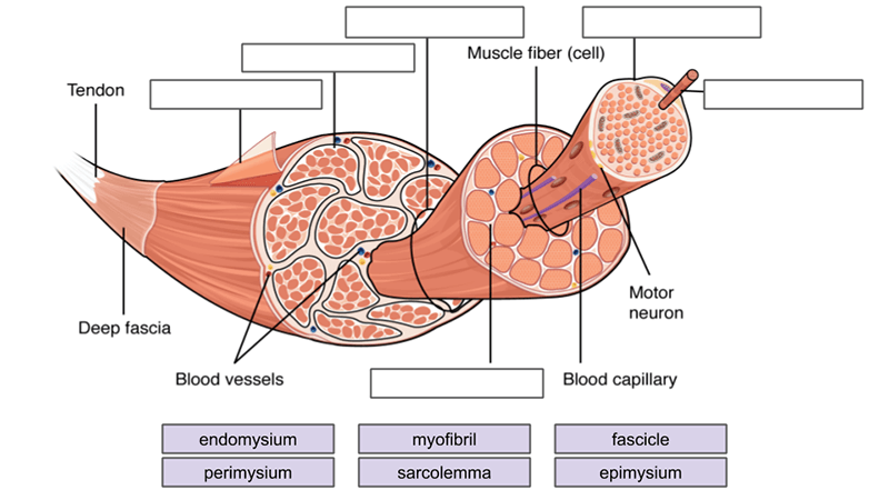
Answered: Art-labeling Activity: Structural… | bartleby Q: Label different areas of an individual muscle unit known as a sarcomere below: Actin A Band Mine… A: The smallest functional unit of muscle tissue is called sarcomere. It consist of actin and myosin…
A & P Ch 6 Musclular System Student PPT
dokumen.pub › campbell-biology-12th-edition-12Campbell Biology, 12th Edition [12nbsped.] 9780135988046 ... 35 Vascular Plant Structure, Growth, and Development Concept 35.1 Plants have a hierarchical organization consisting of organs, tissues, and cells Vascular Plant Organs: Roots, Stems, and Leaves Dermal, Vascular, and Ground Tissues Common Types of Plant Cells Concept 35.2 Different meristems generate new cells for primary and secondary growth
Skeletal muscle tissue: Histology | Kenhub
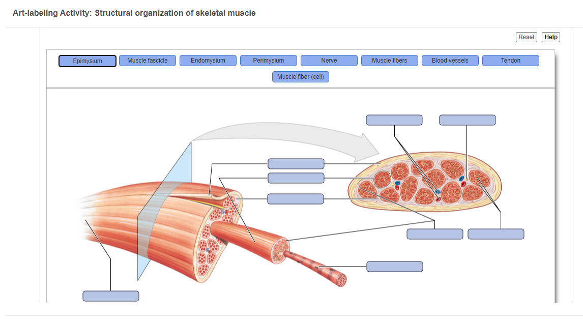
SOLVED: 'Art-labeling Activity: Structural organization of ... - Numerade VIDEO ANSWER:For everyone, so we need to label the figure of the muscle organization right. So this is the organization of muscle, so here at the start we have the connective tissue sheath all right, so this portion over here this is the connective tissue sheet connective tissue sheet. Right now, after the connective tissue sheath, here this portion of the connective tissue sheath, it comes ...

PPT - 9 PowerPoint Presentation, free download - ID:2272780
Solved Secure https:/ C Lab: Histology Art-labeling | Chegg.com Expert Answer. 88% (32 ratings) Answer A is skeletal mucle tissues consisting of = ENDO …. View the full answer. Transcribed image text: Secure https:/ C Lab: Histology Art-labeling Activity: Structure of muscle tissues 102091378 Part A Drag the appropriate labels to their respective targets tissue Smooth muSECM) (cardiac tissue (ECM ...
A&P Chapter 4 Tissue: The Living Fabric Flashcards - Easy ...
AP 1- CHAPTER 9 MASTERING ASSIGNMENTS Flashcards | Quizlet PICTURE Art-labeling Activity: The structure of a skeletal muscle fiber PICTURE Which thin filament-associated protein binds two calcium ions? troponin Action potential propagation in a skeletal muscle fiber ceases when acetylcholine is removed from the synaptic cleft.
Nervous Tissue
quizlet.com › 440072871 › biology-2301-exam-1-studyBIOLOGY 2301 EXAM 1 - STUDY SET CHAPTER, 1, 2, 3, 4 ... Primary structure - sequence of a chain of amino acids secondary structure - hydrogen bonding of the peptide backbone causes the amino acids to fold into a repeating pattern. Tertiary structure - three- dimensional folding pattern of a protein due to side chain interactions.
Skeletal muscle - Wikipedia
9+ art-labeling activity: structure of compact bone most standard Lowest rating: 1. Descriptions: Compact bone is the denser, stronger of the two types of osseous tissue (Figure 6.3.6). It makes up the outer cortex of all bones and is in immediate contact …. More : Compact bone is the denser, stronger of the two types of osseous tissue (Figure 6.3.6).
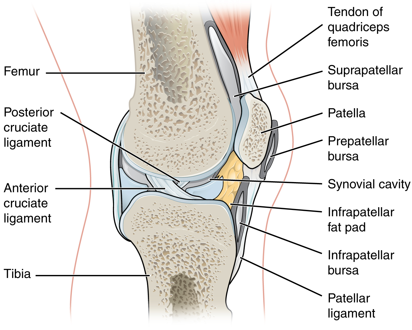
Synovial Joints – Anatomy & Physiology
Mastering AP Chapter 6 - Bones and Skeletal Tissues Study with Quizlet and memorize flashcards containing terms like Art-labeling Activity: Figure 6.2 Long bone Short bone Irregular bone Flat bone Sesamoid bone (short), Art-labeling Activity: Figure 6.4a Distal epiphysis Diaphysis Medullary cavity Compact bone Articular cartilage Proximal Epiphysis Spongy bone Epiphyseal line, Art-labeling Activity: Figure 6.4c Yellow bone …
Answered: Art-labeling Activity: Structural… | bartleby
quizlet.com › 488100546 › chapter-3-lab-3-masteringChapter 3: Lab 3 Mastering Assignment Questions - Quizlet When your biceps brachii (upper arm) muscle contracts, ultimately and most directly, what is producing the movement? 1. You move your arm. 2. Proteins within the cells of the biceps brachii slide past each other lengthwise, shortening the muscle. 3. Your biceps brachii shortens. 4. Your brain tells your muscle to contract.
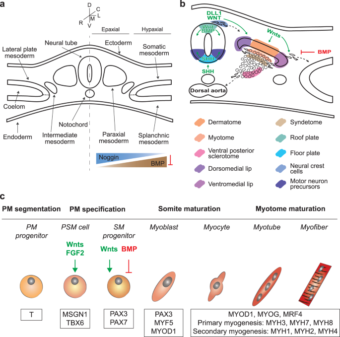
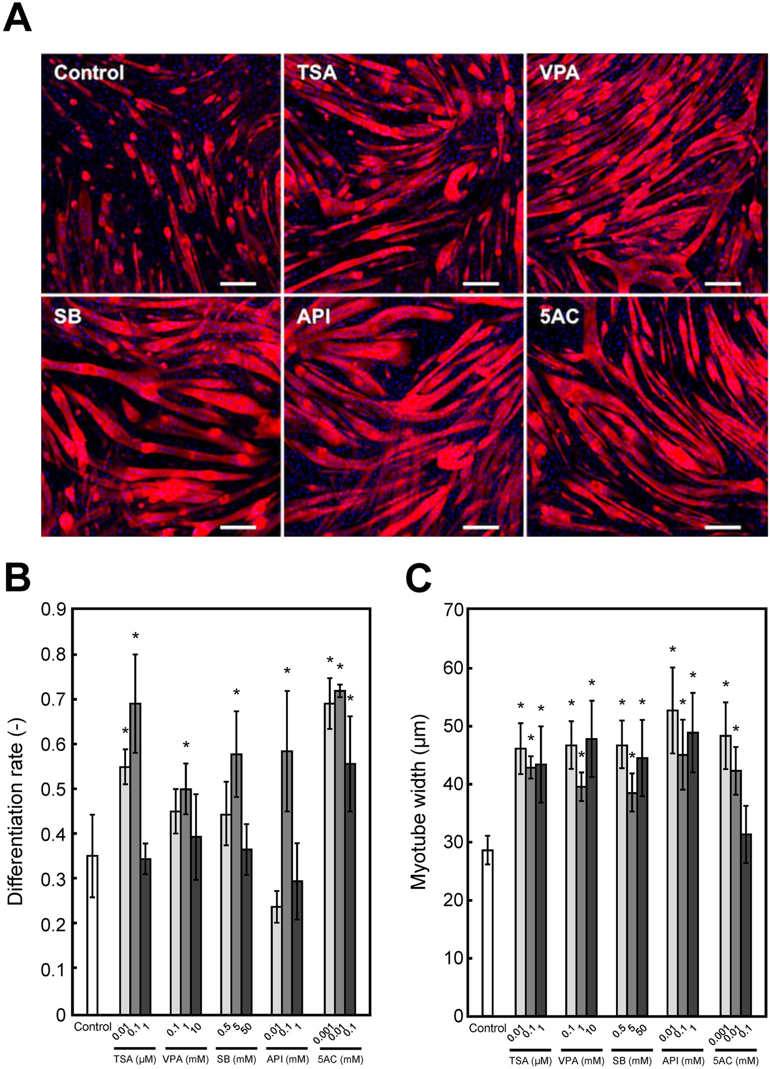
Skeletal muscle differentiation of human iPSCs meets ...
dokumen.pub › human-physiology-an-integratedHuman physiology : an integrated approach [Eighth edition ... Cell Junctions Hold Cells Together to Form Tissues Epithelia Provide Protection and Regulate Exchange Connective Tissues Provide Support and Barriers Muscle and Neural Tissues Are Excitable Tissue Remodeling Apoptosis Is a Tidy Form of Cell Death Stem Cells Can Create New Specialized Cells Emerging Concepts Induced Pluripotent Stems Cells Focus ...
THE MUSCULAR SYSTEM
[Solved] Art-Labeling Activity: | Course Hero All tutors are evaluated by Course Hero as an expert in their subject area. The image attached below is a sketch of the layers of the skin. There are 5 layers of the epidermis, which are categorized as follows (from superficial to deep): Stratum corneum: outer layer of the epidermis. Consists of dead (anuclear), keratin-filled cells.
In vitro drug testing based on contractile activity of C2C12 ...

Exercise 4 Review Sheet Art labeling Activity 1 1 of 2.png ...
Classification of Tissues
Ch. 7

Bioengineered in vitro skeletal muscles as new tools for ...
Muscles and Muscle Tissue

art labeling activity -structure of a nail (superficial and ...
What Do You Know About Bone Physiology , Formation ...
Skeletal muscle - Wikipedia

A & P Ch 6 Musclular System Student PPT
The Muscular System The Muscular System
Chapter 3 Homework - Attempt 1 Art-labeling | Chegg.com
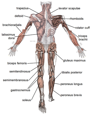
Muscles Labeling
Muscle | histology
A Functional Approach to Human Anatomy Available in a ...

Skeletal muscle heme oxygenase-1 activity regulates aerobic ...
Muscular System Labeling Teaching Resources | Teachers Pay ...
9.17: Skeletal Muscle - Medicine LibreTexts
What does luminal mean in anatomy? - Quora
Bone and Cartilage
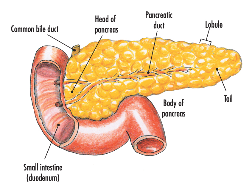
The pancreas | Anatomy of the pancreas | Structure of the ...
Anatomy Review Skeletal Muscle Tissue - Anatomy Review ...
BSCI338N: Diseases of the Nervous System http://www.dartmouth ...
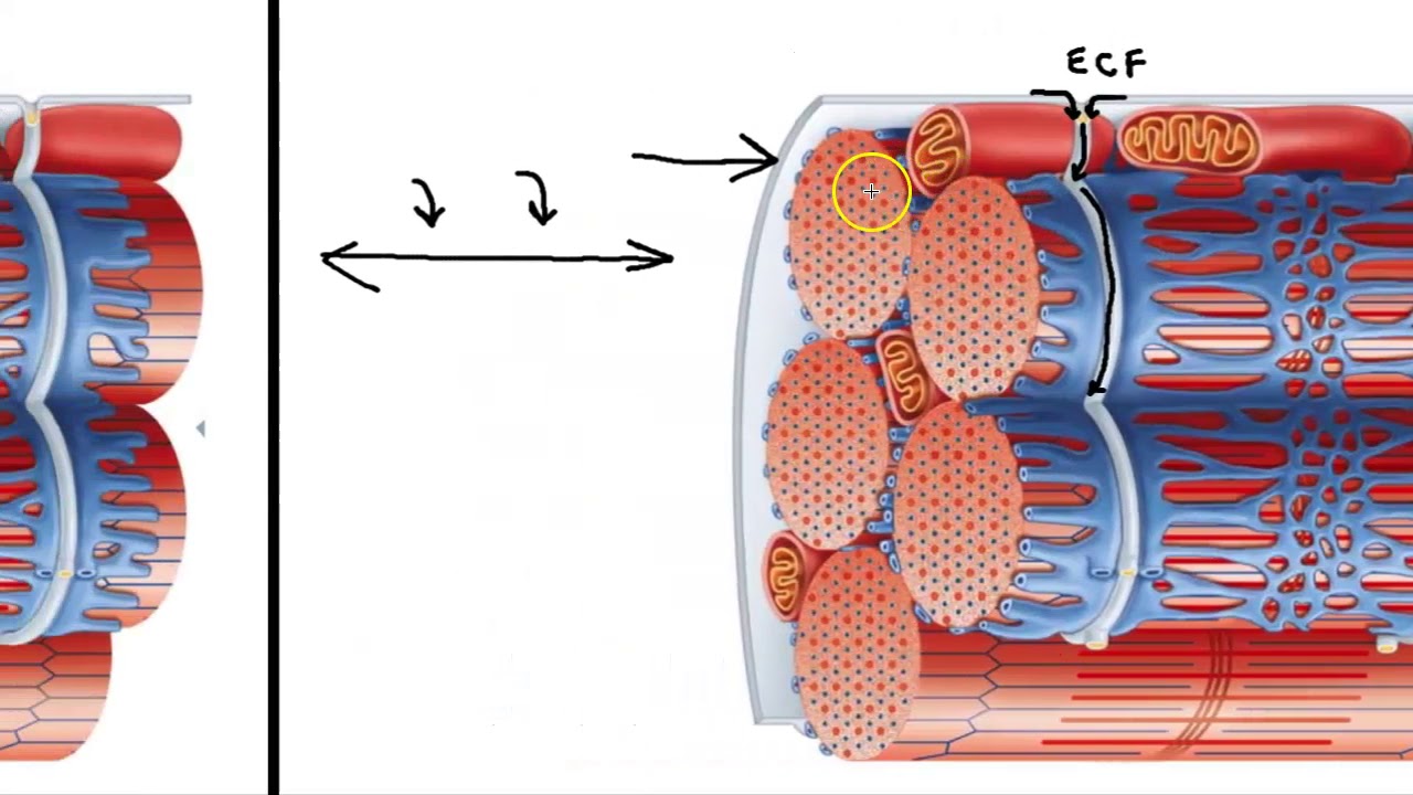
Sarcoplasmic Reticulum and T Tubules

Chapter 9: Muscles and Muscles Tissue | Anatomy and ...
Learn the Essential What, How & Why of Human Anatomy & Physiology
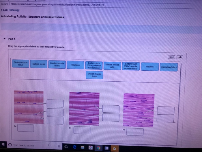
Solved Secure https:/ C Lab: Histology Art-labeling | Chegg.com
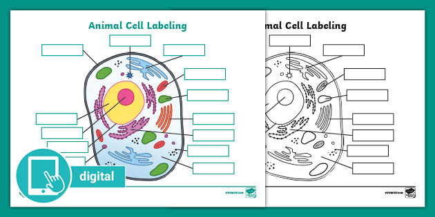
What is an Animal Cell? | Definition and Functions | Twinkl

BIO 200 Chapter 9 - Muscle Tissue Physiology Flashcards | Quizlet

VIDEO LECTURES MENU — Downeast Emergency Medicine
The Muscular System The Muscular System
Post a Comment for "43 art-labeling activity: structure of muscle tissues"